Before treatment
Planning and preliminary examination
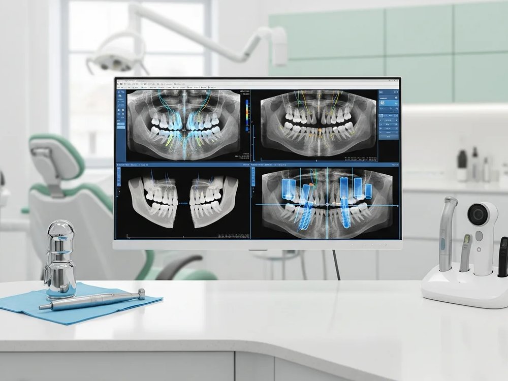
A radiograph gives a first impression of the anatomy, often taken with digital equipment. As far as possible, every potential risk has to be identified before treatment. In implant treatment, diagnostics have a decisive influence on the outcome. Information about the state of the bone at the implant site plays a particularly important part. Is there enough bone, and is its quality sufficient? Bone volume, width and height determine the number of implants. Which implant type, which surface and which length and diameter suit the task? Radiology supplies two-dimensional images with information on the general state of the teeth and the bone.
In more difficult situations these two-dimensional panoramic images are often not enough for reliable implant planning, because the bone can only be assessed to a limited extent. The spatial picture of the bone is missing and the implant site cannot be shown vertically. That carries a risk of injuring anatomical structures such as neighbouring tooth roots, nerves or the maxillary sinus.