Cone beam computed tomography supports diagnosis in critical situations
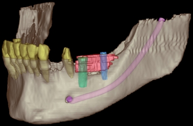
Cone beam computed tomography is a further development of computed tomography. It allows the dental and facial anatomy to be captured three-dimensionally. A single scan can show different viewing angles - axial, coronal and sagittal. The smallest details of the bony facial skull can be visualised and analysed exactly. Cone beam computed tomography produces slice images from a series of X-ray generated pictures. These allow a wide variety of views on the computer. The three-dimensional, plastic representation supports diagnosis, planning and placement of implants in critical situations. In pre-operative implant diagnostics, findings can be identified that are often not visible with conventional imaging techniques. The course of the nerve in the lower jaw, the floor of the maxillary sinus in the upper jaw and the roots of the adjacent teeth can be depicted exactly. The advantage of cone beam computed tomography is a considerably lower radiation dose than with conventional computed tomography, combined with very high resolution.